| |
Practical works are essential for the
understanding of histology and pathology lectures. They
illustrate the true nature of tissues.
The use of a microscope closely relates to the diagnostic
pathway in medicine. During this exercise, the student is
looking for and is gathering various pictures ("symptoms")
from an histological slide, leading to the accurate
diagnosis of the tissue.
The maintenance of a teaching dedicated histology/pathology lab is
very expensive. The constitution of a basic tissue slides
library does take many years. Under some tropical
conditions, microscopes have a very short life expectancy.
These three considerations explain the huge problems
encountered by academics in emerging countries for
establishing such histology labs. This is why we decided to build a
digital microscope which could simulate (although not replace)
the histology/pathology laboratory exercises. The interface of the
digital microscope should allow the user to zoom in and out
in the digital slides and also to move around the slide.
Like an actual microscope...
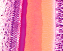
A complete collection of histological and pathological slides, gathered by
Professor Robert Leloup along a 35-year career at
UNamur (formerly FUNDP),
has been scanned at very high resolution (Aperio
technologies). These digital slides are yelding gigabytes
files. Jpeg compression and
Zoomify visualization tools
allow to view these slides from an Internet server. Since
most emerging countries do not benefit from broadband, the
digital microscope will be installed on local servers in
academic institutions.
As a side effect, what is of benefit for students from emerging
countries should be of benefit for our students too...
This project has been supported by
CUD for the scanning of the
slides (TRIBVN).
|
|