The digital microscope has been developed by Prof. Michel Hérin at the University of Namur under the auspices and with the financial support of the Belgian Development Cooperation and the CUD. The digital microscope is aimed at providing high educational material for Medical Faculties localized in emerging countries. It will be installed on internal servers in cooperating institutions all over the world. Moreover, it is currently accessible to everyone from this website.

Practical works are essential for the understanding of histology and pathology lectures. They illustrate the true nature of tissues.
The use of a microscope closely relates to the diagnostic pathway in medicine. During this exercise, the student is looking for and is gathering various pictures ("symptoms") from an histological slide, leading to the accurate diagnosis of the tissue.
The maintenance of a teaching dedicated histology/pathology lab is very expensive. The constitution of a basic tissue slides library does take many years. Under some tropical conditions, microscopes have a very short life expectancy.
These three considerations explain the huge problems encountered by academics in emerging countries for establishing such histology labs. This is why we decided to build a digital microscope which could simulate (although not replace) the histology/pathology laboratory exercises. The interface of the digital microscope should allow the user to zoom in and out in the digital slides and also to move around the slide. Like an actual microscope...
A complete collection of histological and pathological slides, gathered by Professor Robert Leloup along a 35-year career at UNamur (formerly FUNDP), has been scanned at very high resolution (Aperio technologies). These digital slides are yelding gigabytes files. Jpeg compression and Zoomify visualization tools allow to view these slides from an Internet server. Since most emerging countries do not benefit from broadband, the digital microscope will be installed on local servers in academic institutions.
As a side effect, what is of benefit for students from emerging countries should be of benefit for our students too...
This project has been supported by CUD for the scanning of the slides (TRIBVN Healthcare).
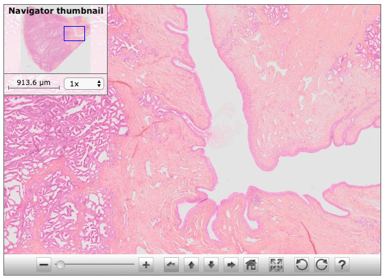
d-microscope Instructions (Zoomify)
April 2020: The d-microscope is now fully HTML5 and responsive (tablet and mobile compatible, included iOS devices). Therefore, it is no longer necessary to install Adobe Flash player.
You should use a broadband Internet access.
Mouse shortcuts:
Alt-click-drag to create a zoom-rectangle.
Alt-click to zoom fully in.
Alt-double-click to zoom fully out.
Alt-click-Reset button to return to the prior view.
(Alt is Option on Macintosh).
The Navigator thumbnail overview can also be clicked or click-dragged to pan.
Use the Toolbar for exact navigation - if using a mouse, hold it over any button to see a helpful tip.
Keyboard shortcuts:
A or Shift to zoom in.
Z or Ctrl to zoom out.
Space Bar to toggle fullscreen view.
Escape to reset initial view or exits fullscreen.
< or > to rotate image if rotation buttons visible.
Short wishlist but huge amount of work:
- A French version of this website (but is this really necessary ?).
- Documented online atlases based on (standstill) pictures extracted from d-microscope Histology and d-microscope Pathology.
- Development of an eWorking platform based on the d-microscopes (for distance training of teachers).
- Collaboration with you... since this site is yours and should be considered as an open platform.